This page was produced as an assignment for Genetics 677, an undergraduate course at UW - Madison.
What are Protein Domains and Motifs?
Protein domains are conserved structural and functional units of a protein. Proteins can be grouped into families based on predicted or known function by knowing which domains the proteins harbor. Domains can confer specific functions that can involve activities such as binding, catalytic capabilities, or signal transduction. While domains refer to tertiary structures of proteins (that is, the physical folding and arrangement of proteins in 3D space), protein motifs refer to only the actual sequence of amino acids [1].
Domains in the PAH Protein
As far as domains go, the PAH protein is pretty simple. The PAH protein contains two domains: an ACT domain and the Biopterin_H domain. ACT domains are involved in binding of small molecules (usually amino acids, such as phenylalanine). In the case of the PAH protein, the ACT domain can undergo a conformational change; this change is relayed throughout the protein and regulates the protein's catabolic function. In typical ACT domain fashion, the domain is located at the end of the protein (in this case, the C-terminal end) [2]. The Bioptern_H domain is a large domain that encompasses almost the entire protein, and binds the cofactor tetrahydrobiopterin (BH4). The domain exists in a family of enzymes that use Iron (II) and function as amino acid hydroxylases. This includes hydroxylases for phenylalanine, tyrosine, and tryptophan [3].

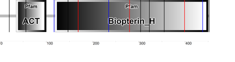
Protein domains as found by Pfam. PAH contains an ACT and Biopterin_H domain.
|
Protein domains as found by SMART. PAH contains an ACT and Biopterin_H domain.
|
The above diagrams were found using two of many programs used to locate protein domains (Pfam and SMART). These programs work by analyzing inputted sequences and comparing them to databases of domains with known sequences. In general, due to the chemical functions and properties of different amino acids, certain positions in the protein are highly conserved in regards to which amino acid (or type of amino acid) is in that particular position. In this sense, if a protein of interest has a sequence that is comparable and similar to a known, conserved region of other proteins, it is likely to have the same function, or domain. Both programs returned comparable results with no detectable discrepancies, that is, both placed an ACT domain at the C-terminus of the protein and a large Biopterin_H domain over the majority of the rest of the protein.
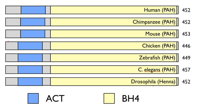
The figure above shows the relative size and location for the ACT and BH4 binding domains for the homologs of the PAH protein. Listed to the right is the protein length in amino acids. It is easy to tell that the PAH protein is highly conserved throughout the animal kingdom - all the homologs have extremely similar sized proteins and the same basic layout and relative sizes of domains. Furthermore there are no additional or truncated domains in any of the homologs. The only possibly notable difference is the ACT domain is closer towards the Biopterin_H domain in humans, chimpanzees, and mice than it is in the rest of the homologs. However, this makes sense, as those three homologs were the closest in sequence and homology in general.
References
1. "Introduction to Protein Classification at the EBI "What Are Protein Domains?" What Are Protein Domains? European Bioinformatics Institute, 2012. Web. 28 Feb. 2013. http://www.ebi.ac.uk/training/online/course/introduction-protein-classification-ebi/protein-classification/what-are-protein-domains
2. Marchler-Bauer A et al. (2013), "CDD: conserved domains and protein three-dimensional structure.", Nucleic Acids Res. 41(D1):D384-52.
From: http://www.ncbi.nlm.nih.gov/Structure/cdd/cddsrv.cgi?uid=214156 [2]
and: http://www.ncbi.nlm.nih.gov/Structure/cdd/cddsrv.cgi?uid=207351 [3]
2. Marchler-Bauer A et al. (2013), "CDD: conserved domains and protein three-dimensional structure.", Nucleic Acids Res. 41(D1):D384-52.
From: http://www.ncbi.nlm.nih.gov/Structure/cdd/cddsrv.cgi?uid=214156 [2]
and: http://www.ncbi.nlm.nih.gov/Structure/cdd/cddsrv.cgi?uid=207351 [3]
